Everything Included Inside the Visual Atlas
A systematic, region-by-region curriculum that bridges the gap between static memorization and real-world functional understanding.
Master exact muscle locations, origins, insertions, actions, innervations, and functional movement chains through an intuitive, streamlined reference system built for effortless recall.
Visual Atlas of Muscle Anatomy — your comprehensive visual study companion to review, test, and reference anytime on your smartphone, tablet, laptop, or printable study sheets.
Progressively structured to help you study faster, retain longer, and quickly reference every key muscle group across the entire human body.

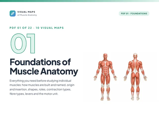
10 visual maps covering how muscles are built and named, origin and insertion, fibre architecture, muscle roles, contraction types, levers and the motor unit.

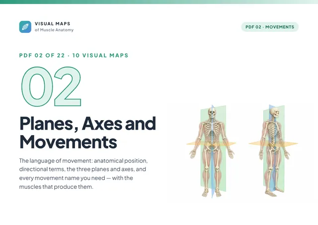
10 visual maps covering anatomical position, directional terms, planes and axes, and every movement: flexion, abduction, rotation, pronation, inversion and more.

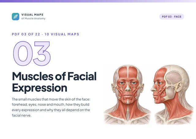
10 visual maps covering occipitofrontalis, orbicularis oculi and oris, the nasal muscles, the smile muscles, the lip depressors, buccinator and platysma.

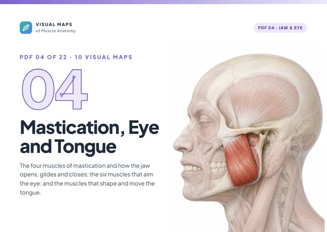
10 visual maps covering masseter, temporalis, both pterygoids, jaw movements, the six extraocular muscles and the muscles of the tongue.

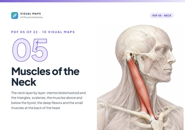
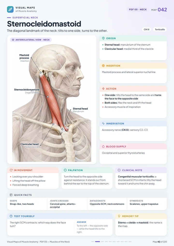
10 visual maps covering the neck triangles, sternocleidomastoid, scalenes, supra- and infrahyoid muscles, deep neck flexors, suboccipitals and splenius.

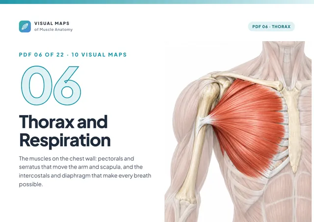
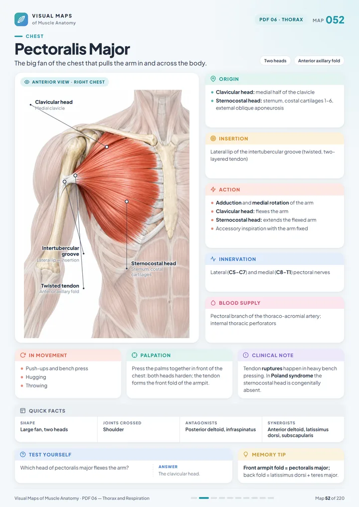
10 visual maps covering pectoralis major and minor, serratus anterior, the intercostals, the diaphragm and the mechanics of breathing.

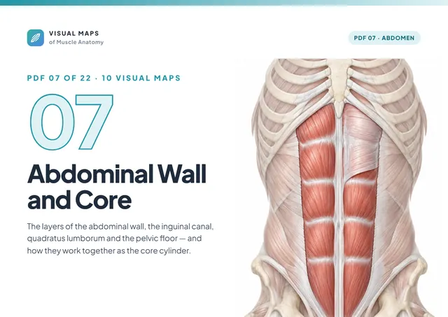
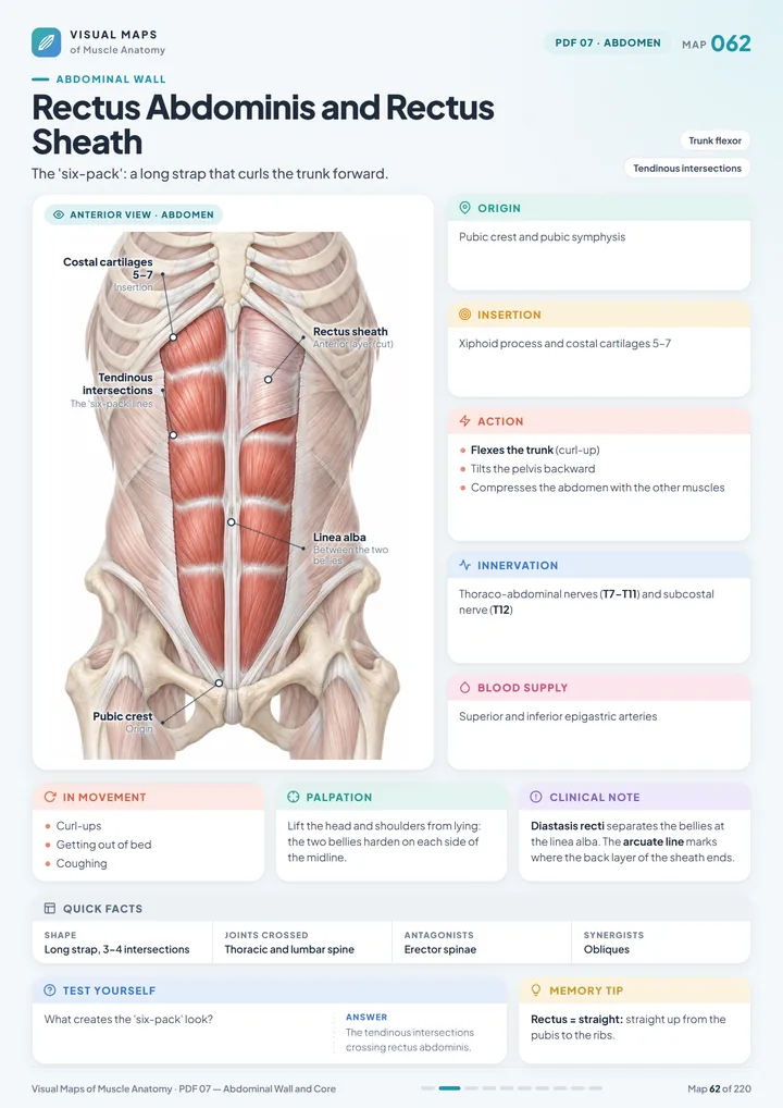
10 visual maps covering rectus abdominis, the obliques, transversus abdominis, the inguinal canal, quadratus lumborum, the pelvic floor and the core.

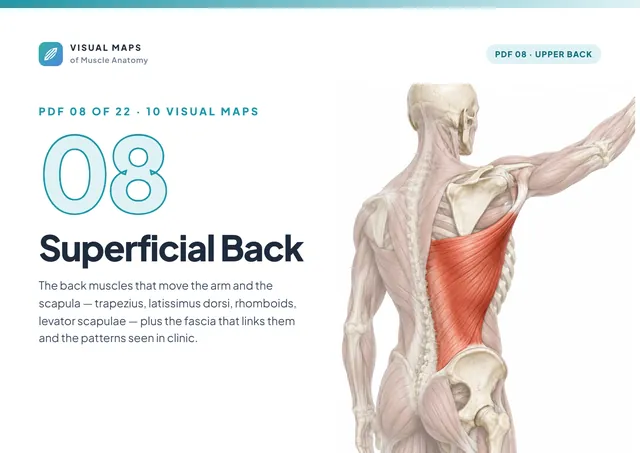
10 visual maps covering trapezius, latissimus dorsi, rhomboids, levator scapulae, the thoracolumbar fascia, scapular force couples and winging.

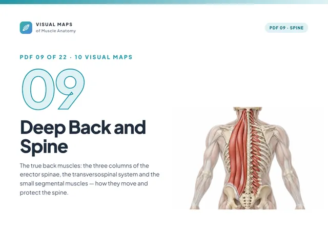
10 visual maps covering iliocostalis, longissimus, spinalis, multifidus, semispinalis, the segmental muscles, trunk movements and lumbar stability.

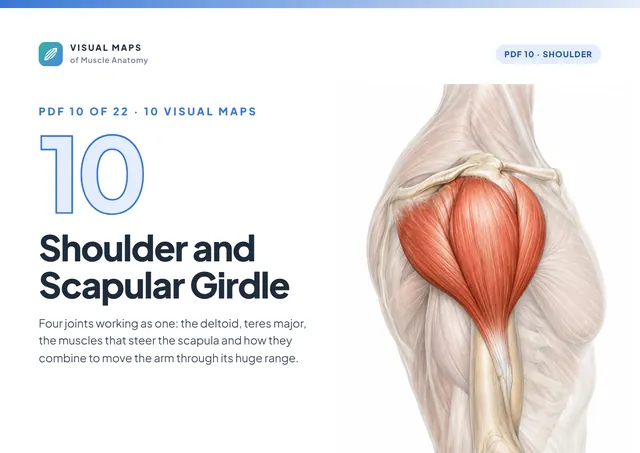
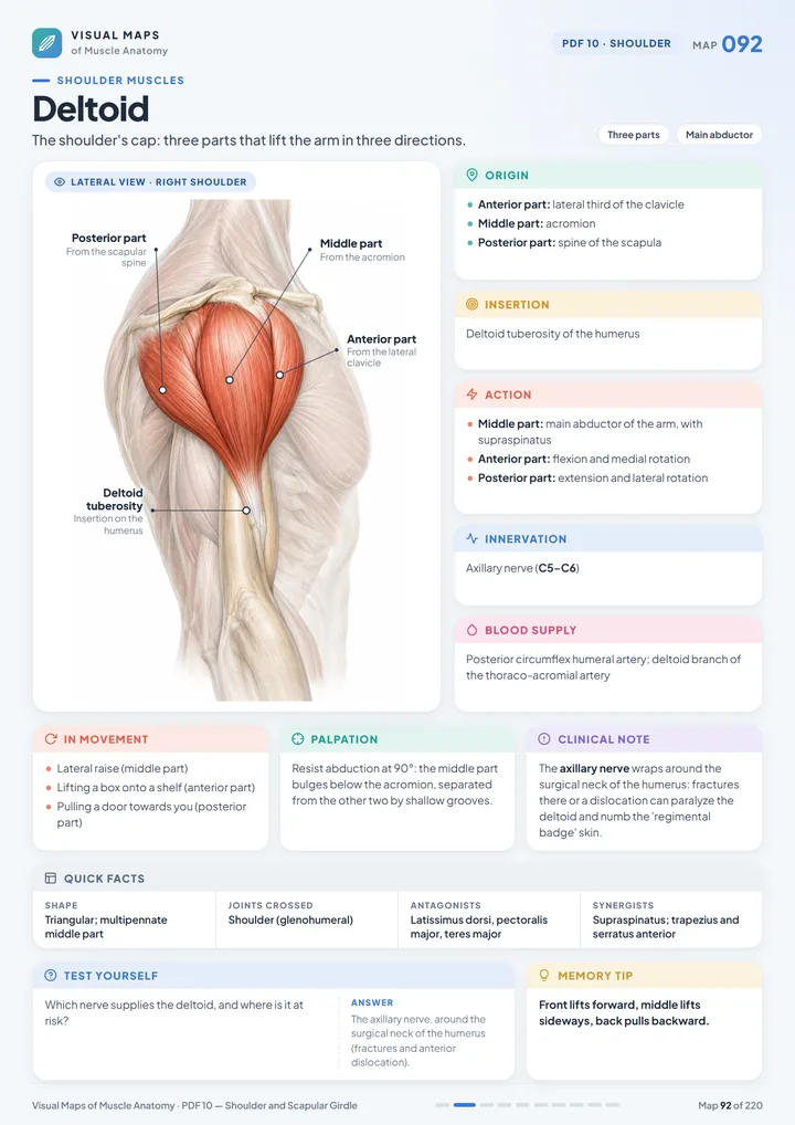
10 visual maps covering the shoulder complex, deltoid, teres major, scapular movements, scapulohumeral rhythm, shoulder movements and impingement.

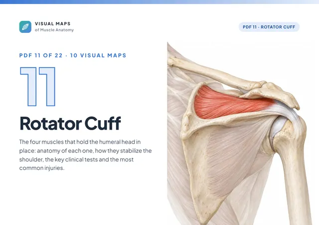
10 visual maps covering supraspinatus, infraspinatus, teres minor, subscapularis, cuff stability, clinical tests, injuries and the posterior spaces.

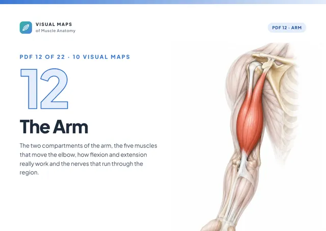
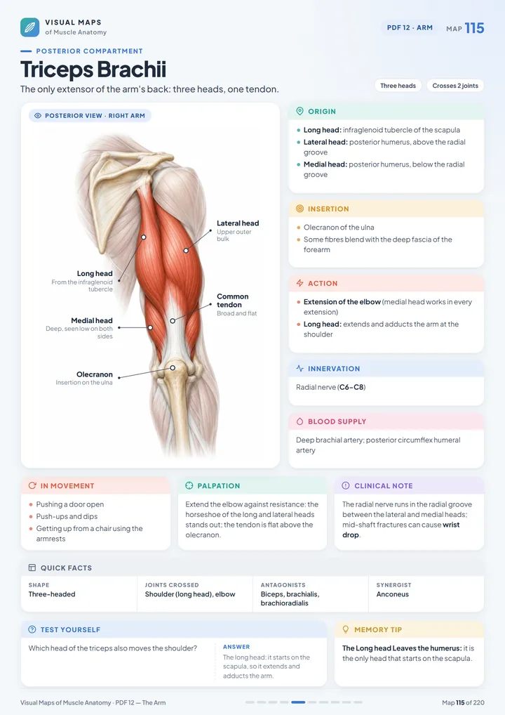
10 visual maps covering the arm compartments, biceps brachii, brachialis, coracobrachialis, triceps, anconeus, elbow flexion and extension and the nerves of the arm.

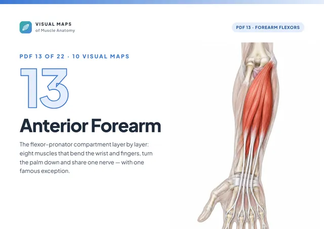
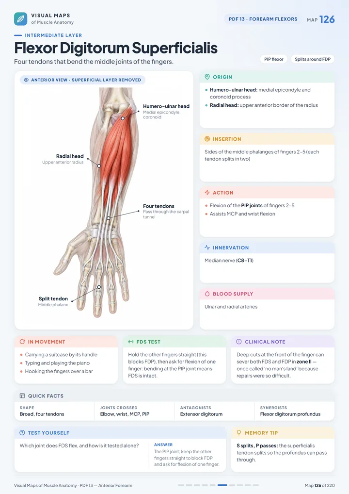
10 visual maps covering pronator teres, the wrist flexors, palmaris longus, flexor digitorum superficialis and profundus, flexor pollicis longus and pronator quadratus.

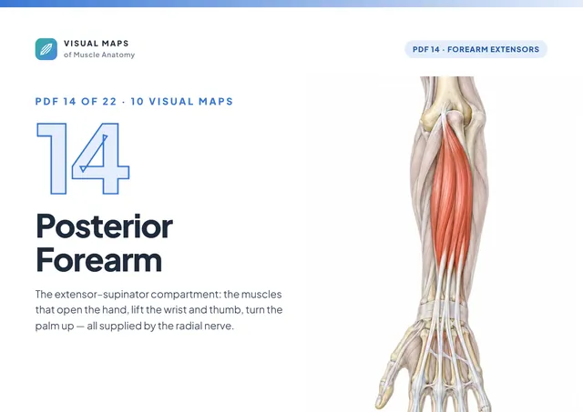
10 visual maps covering brachioradialis, the wrist extensors, extensor digitorum, supinator, the thumb extensors, the anatomical snuffbox and tennis elbow.

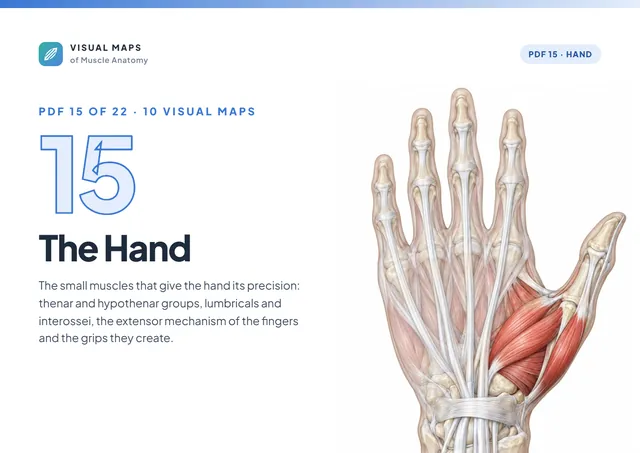
10 visual maps covering the thenar and hypothenar muscles, adductor pollicis, lumbricals, interossei, the extensor mechanism of the fingers and the grips.

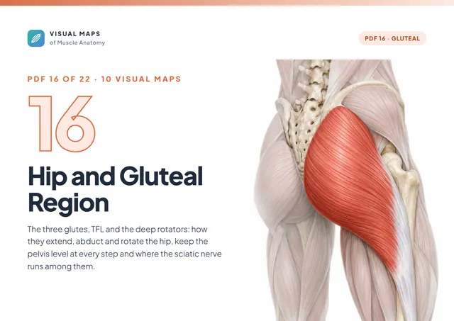
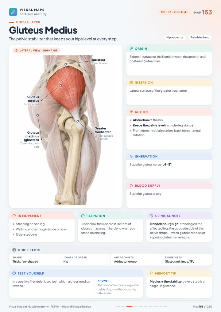
10 visual maps covering the three glutes, tensor fasciae latae and the IT band, piriformis, the deep rotators, the abductors in gait and hip extension.

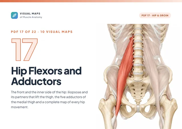
10 visual maps covering psoas major, iliacus, the adductor group, adductor magnus, pectineus, gracilis and a complete map of hip movements.

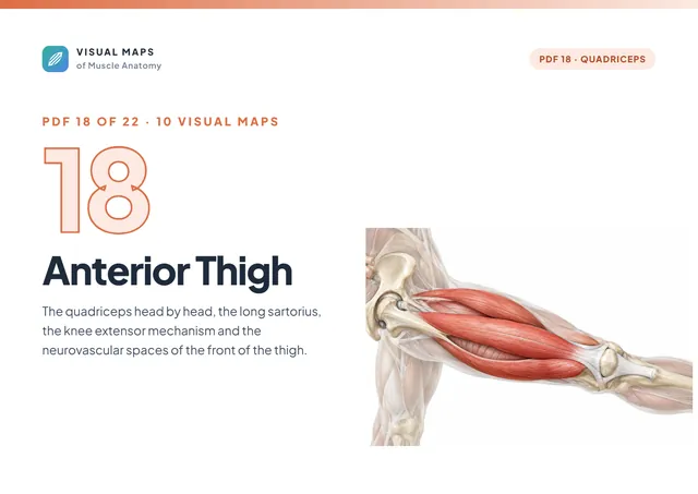
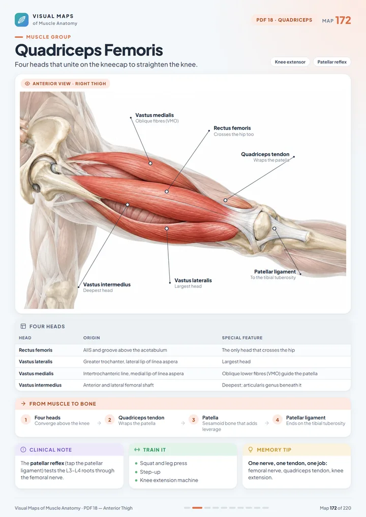
10 visual maps covering the quadriceps head by head, sartorius, the knee extensor mechanism and the femoral triangle.

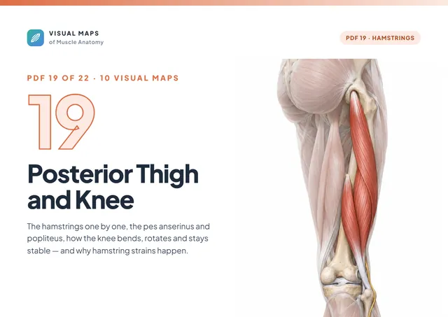
10 visual maps covering biceps femoris, semitendinosus, semimembranosus, the pes anserinus, popliteus, knee movements and hamstring strain.

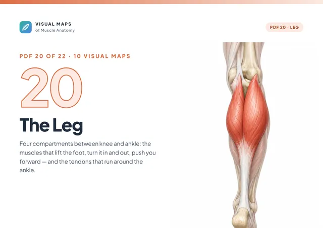
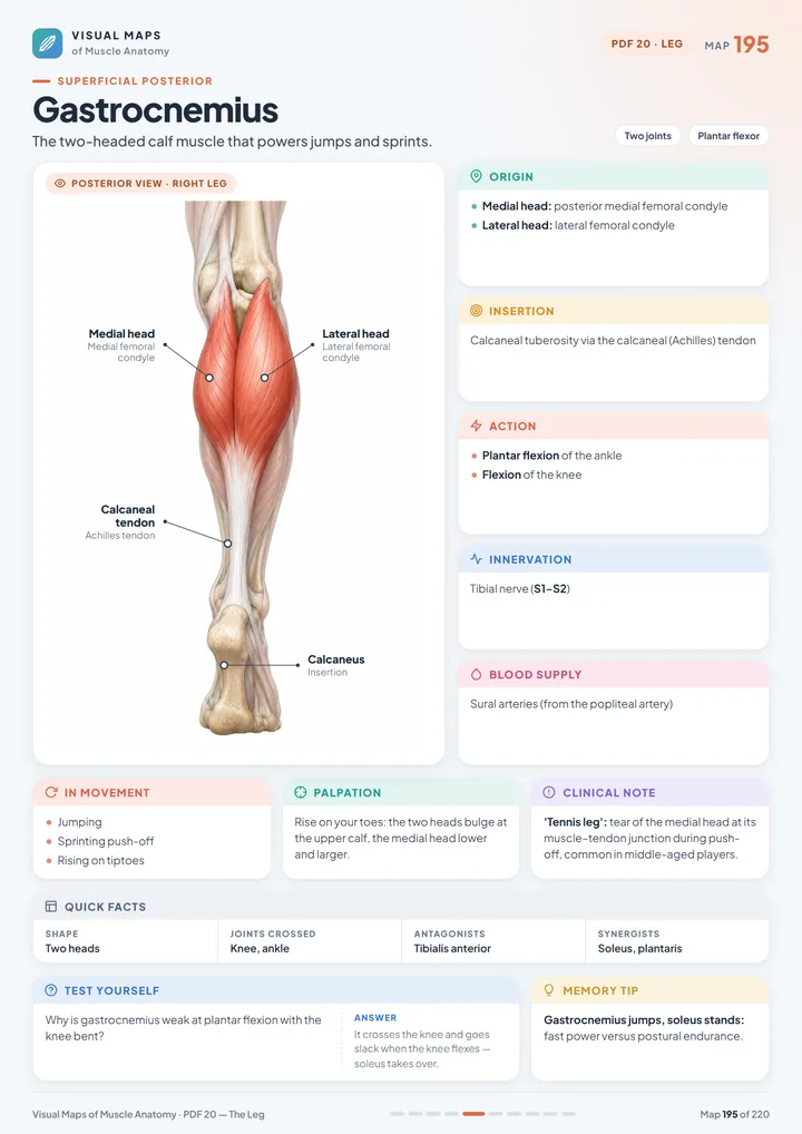
10 visual maps covering the leg compartments, tibialis anterior, the toe extensors, fibularis longus and brevis, gastrocnemius, soleus and tibialis posterior.

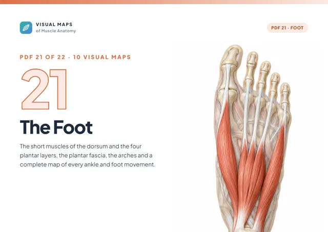
10 visual maps covering the dorsum of the foot, the four plantar layers, the plantar aponeurosis, the arches and every ankle and foot movement.

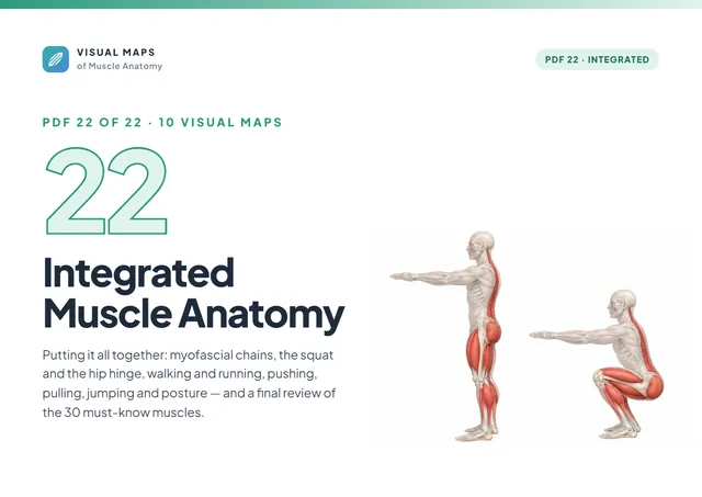
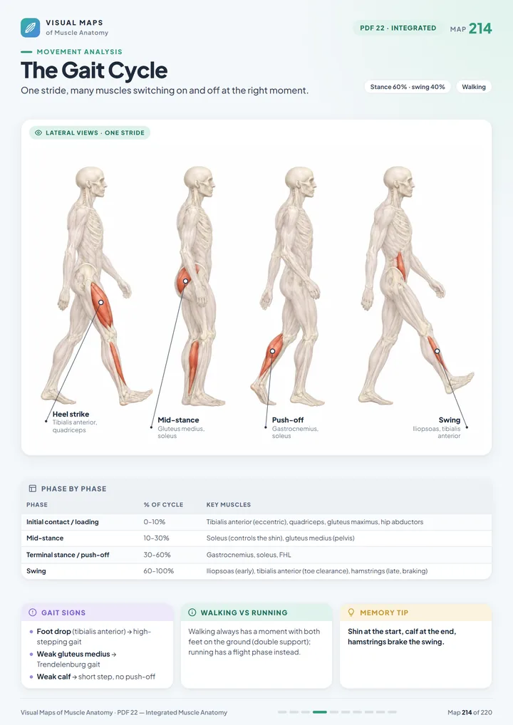
10 visual maps covering myofascial chains, the squat, the hip hinge, gait, running, pushing, pulling, jumping, posture and the 30 must-know muscles.
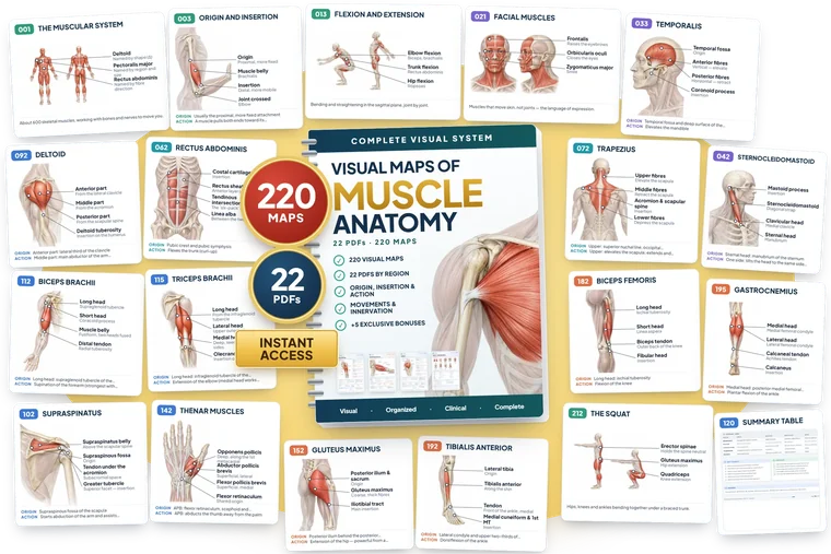
Crystal-clear anatomical illustrations, side-by-side comparative charts, and functional movement schematics that make muscle anatomy click instantly.
The Visual Atlas was crafted for active students and clinicians who need crystal-clear, reliable anatomical recall without wasting hours on dense textbook walls of text.
Get the complete Visual System of Muscle Anatomy, with all 22 PDFs of the main material and everything presented on this page.
Unlock the Complete Atlas Now
A systematic, region-by-region curriculum that bridges the gap between static memorization and real-world functional understanding.
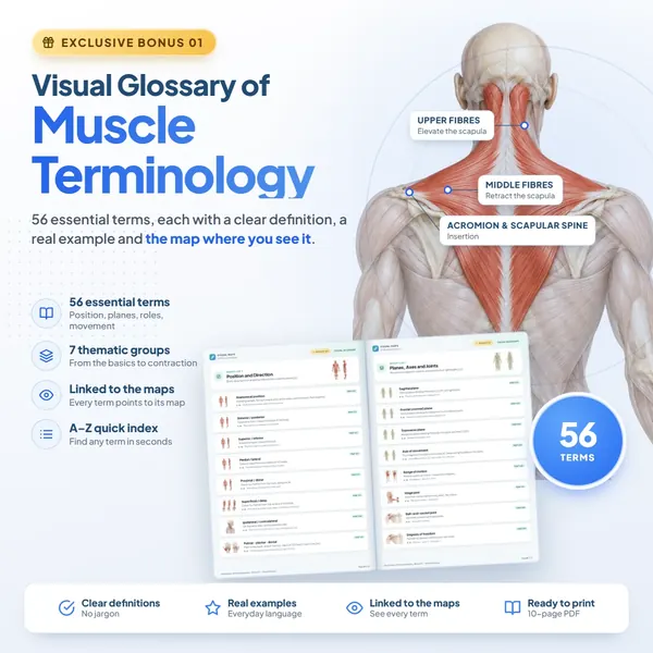
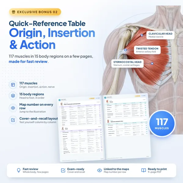
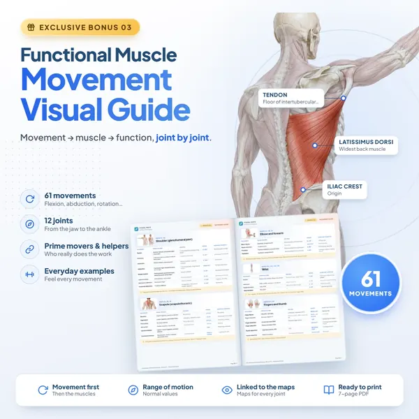
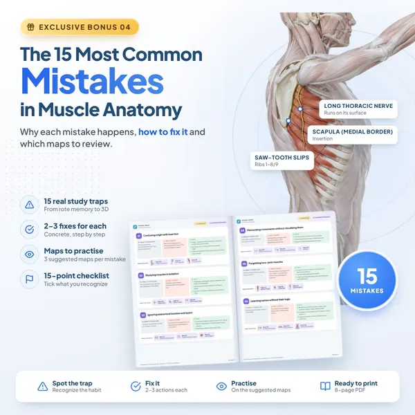
Powerful companion tools designed to decode terminology, accelerate exam prep, and structure your daily review into a friction-free routine.
I always inverted origin and insertion on practical tests. Seeing them laid out side by side on one visual sheet finally fixed that in just a few days of review.
Sarah Jenkins
Physiotherapy student · 5th semester
I reviewed these maps the evening before my practical anatomy bell-ringer. I recognized every muscle on the pinned specimens immediately without second-guessing.
Ethan Reynolds
Medical student · 3rd year
As a coach and trainer, I used to juggle three textbooks just to explain a simple movement chain. Now I pull up the visual guide on my phone and show my clients right away.
Marcus Bennett
Exercise professional
Connecting muscle structure directly with its action and innervation in one clean graphic completely changed how I study. No more memorizing random disconnected lists.
Chloe Henderson
Nursing student
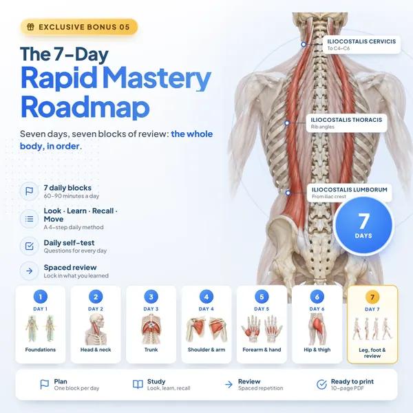
The 7-Day roadmap gave me a real study strategy. Before, I’d open my anatomy atlas with zero direction and get overwhelmed in 20 minutes.
Emily Carter
Biomedicine student
The master origin, insertion, and action table is easily my favorite tool. I can review the entire lower extremity thoroughly in fifteen minutes before clinic.
Tyler Davis
Personal trainer
I'm in Occupational Therapy and the forearm and hand breakdown is a lifesaver. The intrinsic muscle maps are clean, intuitive, and extremely easy to follow.
Hannah Brooks
Occupational Therapy student
I have been lecturing anatomy for eight years. I now recommend these visual sheets to all my freshmen. Student retention in lab exams has noticeably improved.
Prof. Andrew Harrison
Anatomy lecturer
I review on my phone during my transit commute every morning. Since the PDFs are clean and readable, I can conquer an entire muscle compartment per ride.
Jessica Taylor
Physiotherapy student
The 15 common mistakes guide pinpointed my exact blind spot: memorizing muscles isolated from their joint mechanics. My comprehension skyrocketed.
Lucas Morgan
Exercise Science student
I always inverted origin and insertion on practical tests. Seeing them laid out side by side on one visual sheet finally fixed that in just a few days of review.
Sarah Jenkins
Physiotherapy student · 5th semester
I reviewed these maps the evening before my practical anatomy bell-ringer. I recognized every muscle on the pinned specimens immediately without second-guessing.
Ethan Reynolds
Medical student · 3rd year
As a coach and trainer, I used to juggle three textbooks just to explain a simple movement chain. Now I pull up the visual guide on my phone and show my clients right away.
Marcus Bennett
Exercise professional
Connecting muscle structure directly with its action and innervation in one clean graphic completely changed how I study. No more memorizing random disconnected lists.
Chloe Henderson
Nursing student
The 7-Day roadmap gave me a real study strategy. Before, I’d open my anatomy atlas with zero direction and get overwhelmed in 20 minutes.
Emily Carter
Biomedicine student
The master origin, insertion, and action table is easily my favorite tool. I can review the entire lower extremity thoroughly in fifteen minutes before clinic.
Tyler Davis
Personal trainer
I'm in Occupational Therapy and the forearm and hand breakdown is a lifesaver. The intrinsic muscle maps are clean, intuitive, and extremely easy to follow.
Hannah Brooks
Occupational Therapy student
I have been lecturing anatomy for eight years. I now recommend these visual sheets to all my freshmen. Student retention in lab exams has noticeably improved.
Prof. Andrew Harrison
Anatomy lecturer
I review on my phone during my transit commute every morning. Since the PDFs are clean and readable, I can conquer an entire muscle compartment per ride.
Jessica Taylor
Physiotherapy student
The 15 common mistakes guide pinpointed my exact blind spot: memorizing muscles isolated from their joint mechanics. My comprehension skyrocketed.
Lucas Morgan
Exercise Science student
Secure lifetime access to the Visual Atlas today and keep an infallible visual reference ready on all your devices.
Essential access
Pay in your local currency, converted automatically at checkout
Note: this includes the core 220 visual guides. Upgrade to Complete access for all 5 companion bonus guides.
Complete access
Pay in your local currency, converted automatically at checkout
🔒 Secure checkout and instant access·Digital material·Single one-time payment — zero recurring fees
Take 7 full days to explore the Visual Atlas on your phone, tablet, or computer. Test the guides with your coursework, lab exams, or clinical sessions. If you don't feel significantly more confident identifying muscle origins, insertions, and movements, simply request a prompt refund within 7 days — zero questions asked.
Everything you need to know about your access, file formats, and guarantees.
The Visual Atlas is 100% digital and delivered immediately as high-resolution PDF files. No physical book is shipped, meaning zero shipping fees and instant delivery right after checkout.
Immediately after payment confirmation, you will receive an email with your secure login and direct download links to access and save all 12 modules and bonus materials.
You get full lifetime access with no expiration dates and no monthly subscriptions. Once downloaded, the files are yours to keep and reference forever.
You do not need to print them. All PDFs are fully optimized for smooth reading on smartphones, tablets, and computers. However, they are also formatted in high-resolution standard page sizes if you prefer to print physical study binders.
It was engineered for anyone learning or working with human musculoskeletal anatomy: students and practitioners in Medicine, Physical Therapy, Kinesiology, Nursing, Athletic Training, Chiropractic, and Personal Training.
Not at all. Module 01 starts from the ground up with foundational terminology, planes, and movement axes before progressing into specific muscular compartments. It works equally well for beginners and for advanced clinicians refreshing their knowledge.
Yes, in detail. Each muscle guide clearly outlines exact skeletal origin, insertion attachment, primary and secondary actions, and motor innervation in a visual, uncluttered layout.
Absolutely. Thousands of students use these guides specifically for rapid revision before practical bell-ringer exams, anatomy quizzes, and licensing board exams.
No. This is a high-yield visual companion created to accelerate your learning. It takes the dense, confusing descriptions from 1,000-page textbooks and condenses them into clear, memorable visual frameworks.
No. Each purchase grants a single-user personal license. The materials are protected by international copyright laws and are intended strictly for your personal educational use.